


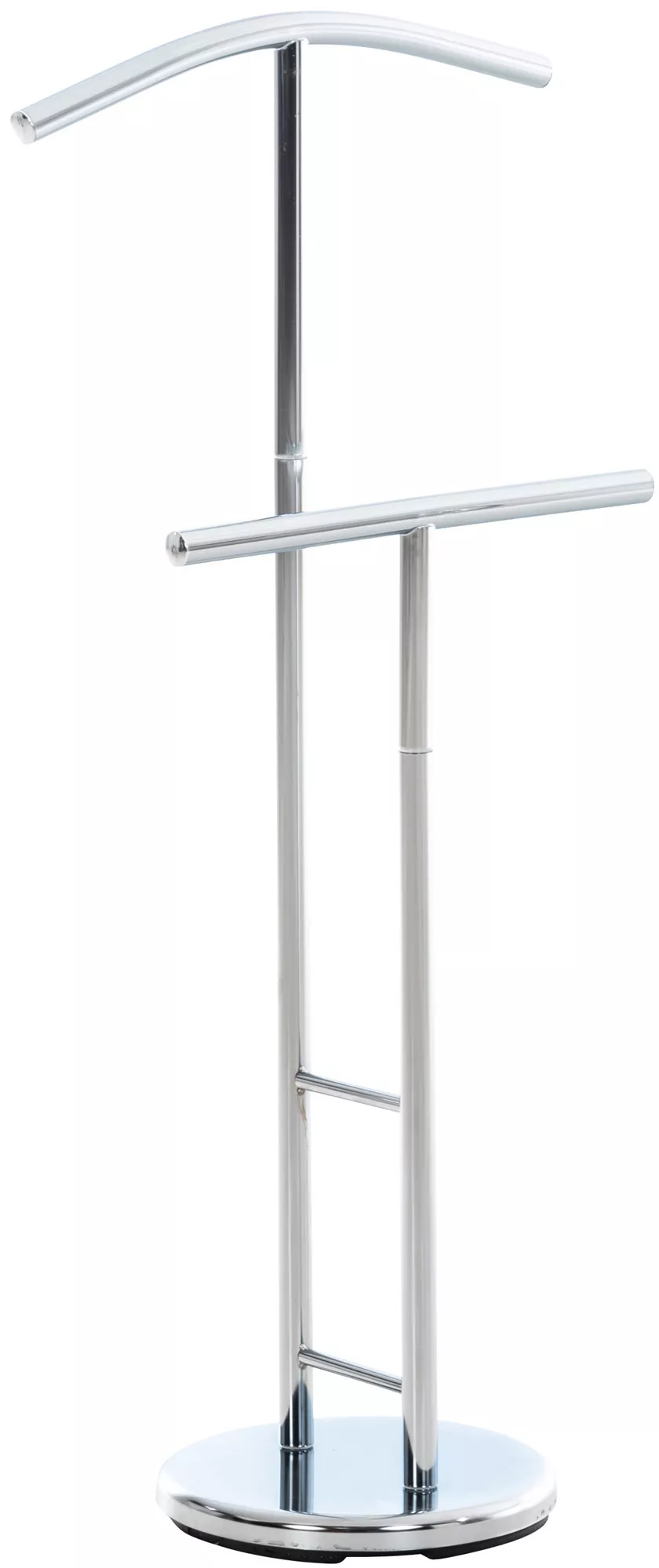
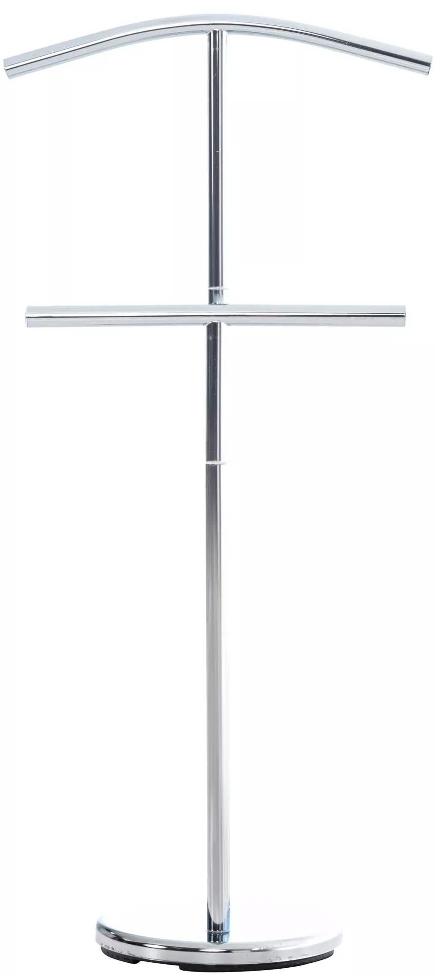

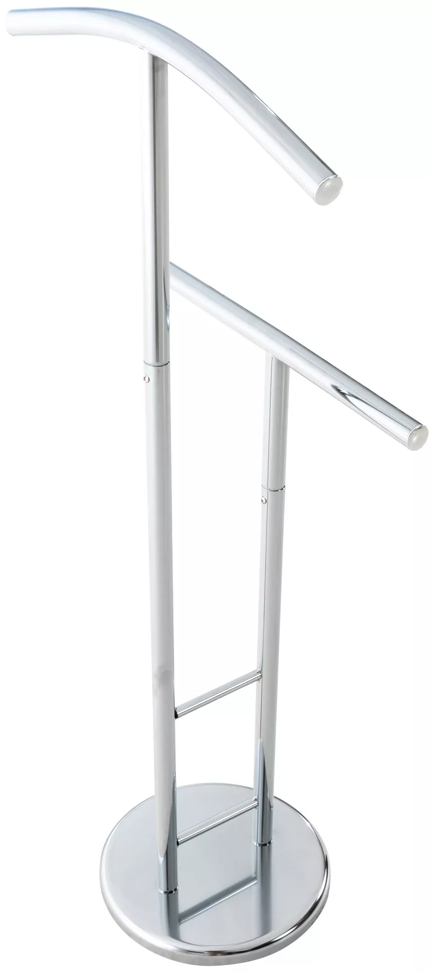
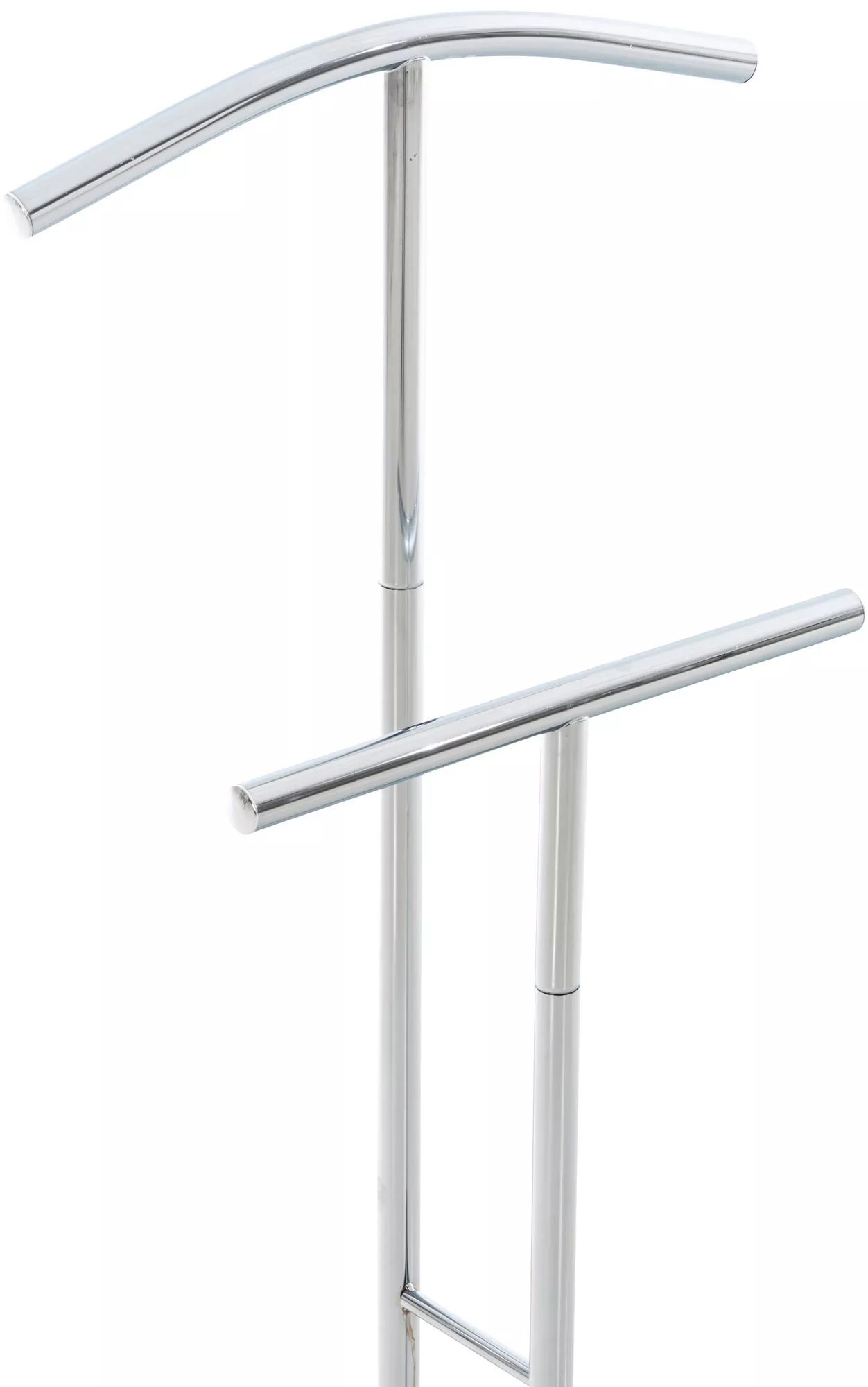

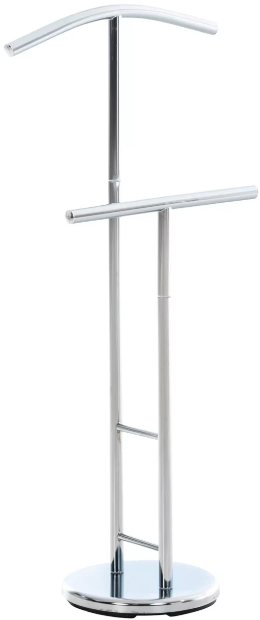


„Raymon valet stand
€119.99
Turime
Drabu
žin
The following table has information from the previous paper (Table 1) and other related data:
| Experiment Parameters | Sample Characteristics | Microscope Specifications | Cell and Nuclear Parameters | Analysis Results | |||||
| Exposure Time | 0.2 sec | Sample Preparation | Paraffin embedding | Microscope Model | Zeiss Axio Imager | Cell Count | 500 | Nuclear Area | 40-60 µm² |
| Magnification | 40x | Staining Method | Hematoxylin and Eosin | Camera Model | Hamamatsu Orca Flash 4.0 | Nuclear Count | 300 | Nuclear Perimeter | 25-35 µm |
| Light Source | LED | Tissue Type | Liver | Software | Zen Blue | Cytoplasmic Area | 70-90 µm² |
Nuclear Circularity | 0.7-0.9 |
| Filter Set | GFP | Species | Mouse | Objective Lens | Plan-Apochromat 40x/1.3 Oil | Cell Shape | Polygonal | Area Ratio (Nuclear/Cytoplasmic) | 0.4-0.6 |
Key Findings and Conclusions: The analysis revealed significant variations in nuclear size and shape within the liver tissue samples. The observed nuclear circularity values suggest a range of nuclear morphology. The area ratio between the nucleus and cytoplasm also shows variability, indicating potential differences in cellular activity or state.
| Svoris | 5 kg |
|---|---|
| Išmatavimai | 28 × 50 × 107 cm |
| Spalva | Sidabras |
| Medžiaga | Metalas |
| Stovo medžiaga | Metalas |
Panašūs produktai
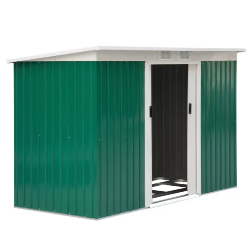

Sodo spintos ir sandėliukai
Metalinis sandėliukas su vėdinimo langu, vienšlaičiu stogu – tamsiai žalia, 280 x 130 x 172 cm


Sodo spintos ir sandėliukai


Sodo spintos ir sandėliukai
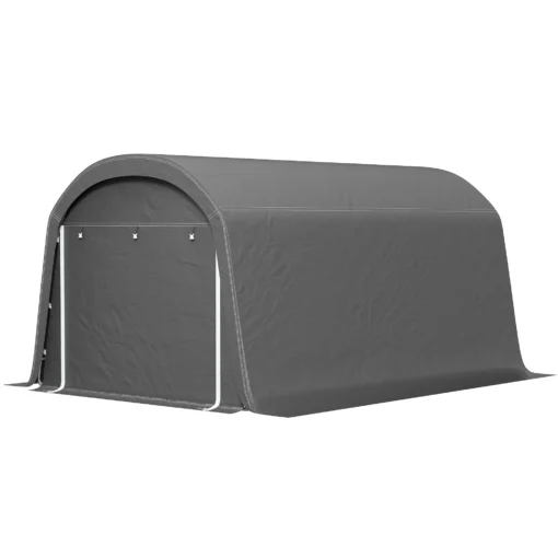

Sodo spintos ir sandėliukai
Vandeniui atspari dviračių stoginė su durimis, apsauga nuo dulkių, pastogė, 300x450x230 cm, pilka


Sodo spintos ir sandėliukai


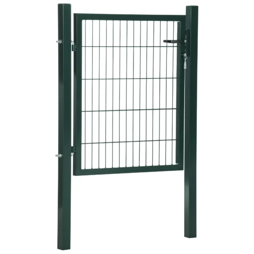
Sodo spintos ir sandėliukai
Sodo įrankių sandėliukas su stumdomomis durimis 277 x 195 x 192 cm

Sodo spintos ir sandėliukai


Sodo spintos ir sandėliukai
Metalinis sandėliukas, įrankių stoginė – šviesiai žalia 277 x 195 x 192 cm
